Views: 0 Author: Site Editor Publish Time: 2026-09-15 Origin: Site
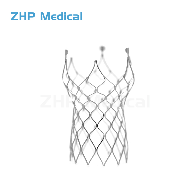
Treating complex vascular lesions in dynamic environments like the superficial femoral artery (SFA) and popliteal artery presents a severe biomechanical challenge. In these anatomical zones, continuous external compression, torsion, and vessel flexion cause traditional rigid stents to fracture, kink, or fail prematurely. Vascular specialists require a robust scaffold that maintains lumen patency, adapts to relentless mechanical stress, and minimizes vessel wall trauma without succumbing to structural fatigue. The self expanding nitinol stent serves as the standard-of-care solution for these hostile environments. Its unique material properties, specifically superelasticity, combined with advanced manufacturing processes and specialized deployment mechanics, dictate clinical outcomes and device selection. We rely on these devices to navigate tortuous anatomy where balloon-expandable options fall short. Understanding how these scaffolds function in vivo helps operators optimize procedural success, mitigate risks, and ensure long-term vessel health for patients.
Material Superiority: Nitinol’s superelasticity allows the stent to recover from large strains and severe mechanical deformation, making it ideal for peripheral arteries subject to bending and compression.
Biomechanical Balance: Once deployed, these stents apply a continuous, low "chronic outward force" (COF) against the vessel wall while providing a high radial resistive force (RRF) against external compression.
Deployment Mechanism: Unlike balloon-expandable variants, a Self-Expanding Nitinol Stent is constrained within a delivery catheter and opens autonomously upon release, requiring specific operator handling techniques to ensure precise placement.
Design-to-Outcome Correlation: Strut thickness, surface friction, and scaffolding design directly influence the rate of vessel injury, endothelialization speed, and subsequent neointimal hyperplasia (restenosis).
Table of Contents
Nitinol is a near-equiatomic nickel-titanium alloy that can reversibly transform between rigid austenite and flexible martensite phases. In a Self-Expanding Nitinol Stent, the Af temperature is typically tuned below body temperature so the stent remains austenitic at 37°C and maintains its expanded shape. When compressed into a delivery catheter, stress transforms the material into martensite, allowing significant deformation without permanent damage. Once released, it returns to austenite and recovers its original shape. Unlike stainless steel or cobalt-chromium stents that rely on permanent plastic deformation, Nitinol can absorb external strain and rebound, making it well suited for peripheral arteries exposed to continuous movement.
Manufacturing a Nitinol vascular scaffold requires precise control of geometry, heat treatment, and surface quality. The stent is laser-cut from Nitinol tubing, often using femtosecond lasers to minimize heat damage and micro-cracking. It then undergoes heat setting at approximately 450–550°C to program its final shape. After cutting and heat treatment, electropolishing removes surface oxides and irregularities while creating a smooth, passivated titanium dioxide (TiO₂) layer. This smoother surface reduces friction and platelet adhesion while supporting endothelialization, which is important for long-term vessel healing.
Peripheral arteries, particularly the SFA and the segment passing through Hunter's canal, are subjected to a brutal, unyielding biomechanical environment. With every step a patient takes, these vessels undergo complex, multi-directional forces. A rigid metallic stent placed in this dynamic zone will quickly experience metal fatigue, leading to catastrophic strut fracture and subsequent vessel occlusion.
We categorize the primary forces acting on these stents into four distinct types:
Torsion: Twisting motions caused by the rotation of the leg and knee joint.
Flexion: Severe bending, particularly behind the knee during walking or sitting.
Extension: Longitudinal stretching of the artery as the limb extends.
Compression: Direct external crushing forces from surrounding skeletal musculature contracting against the vessel.
The superelastic properties of nitinol allow the stent to absorb these multi-directional forces effectively. The material can accommodate up to 8% recoverable strain without suffering permanent plastic deformation or structural failure. To ensure the design can withstand millions of gait cycles over a patient's lifetime, engineers rely heavily on Finite Element Analysis (FEA). FEA computational modeling simulates the exact physical forces the stent will face in vivo. This allows designers to accurately predict fatigue life, identify high-stress concentration points at specific strut junctions, and optimize the overall geometry before clinical manufacturing and human trials begin.
The clinical success and long-term patency of a self-expanding scaffold hinge on the delicate balance between two distinct mechanical forces: Chronic Outward Force (COF) and Radial Resistive Force (RRF). Nitinol exhibits a unique biomechanical property known as the hysteresis loop. When plotting stress against strain, the force required to compress the stent (the loading plateau) is significantly higher than the force the stent exerts as it expands (the unloading plateau). This hysteresis is clinically vital.
Biomechanical Force | Definition | Clinical Function | Ideal Target Profile |
|---|---|---|---|
Chronic Outward Force (COF) | The continuous, gentle outward pressure the stent exerts against the arterial wall after deployment. | Maintains lumen patency and ensures the stent stays apposed to the vessel wall over time. | Low. A low COF prevents excessive mechanical trauma to the intima, reducing the inflammatory response and limiting neointimal hyperplasia. |
Radial Resistive Force (RRF) | The structural ability of the stent to resist external compressive forces. | Prevents the vessel from collapsing under the pressure of muscle contractions or external trauma. | High. A high RRF ensures the scaffold can hold the artery open even in heavily diseased or compressed anatomical zones. |
This hysteresis loop allows the stent to provide a robust, high RRF to prevent the vessel from collapsing under external pressure, while simultaneously maintaining a low COF. If a stent design fails to achieve this balance and exerts a high COF, it will cause deep intimal injury. The physiological response to this continuous barotrauma is rapid smooth muscle cell proliferation, leading to severe intimal hyperplasia and eventual restenosis.
Human arteries are not static, rigid pipes. They are dynamic, pulsatile, and naturally tapered structures. A self-expanding scaffold adapts to these anatomical variations. As the artery expands and contracts with each systolic and diastolic heartbeat, the flexible nitinol structure flexes in unison with the vessel wall. Because the stent is intentionally oversized relative to the target vessel diameter (typically by 1 to 2 millimeters), it maintains constant, secure apposition against the intima, even in highly tapered arterial segments.
Optimal sizing is a strict procedural parameter. If an operator aggressively oversizes the stent (e.g., placing an 8mm stent in a 5mm vessel), the resulting high COF stretches the medial layer of the artery. Proper sizing balances the mechanical need for secure wall apposition and migration resistance against the biological risk of mechanically induced inflammation. We rely on precise intravascular ultrasound (IVUS) or quantitative vessel angiography to determine the exact reference vessel diameter before selecting the stent size.
Unlike balloon-expandable stents that are crimped onto a deflated balloon, a self-expanding device is constrained within a specialized, multi-layered delivery catheter. The architecture of this delivery system typically includes a flexible inner shaft that holds the stent in place and an outer constraining sheath that tightly compresses the nitinol structure. Radiopaque markers made of tantalum or platinum-iridium are positioned on both the inner shaft and the stent ends. These markers allow the operator to visualize the device clearly under fluoroscopy.
Deployment occurs when the operator physically retracts the outer sheath. As the mechanical constraint is removed, the nitinol scaffold autonomously expands to its pre-set diameter, anchoring itself to the vessel wall. Historically, delivery systems utilized a simple "pin-and-pull" mechanism, requiring the operator to manually pull back the outer sheath while attempting to hold the inner shaft perfectly steady. Modern systems now utilize advanced micro-geared or thumb-wheel handles. These ergonomic, mechanical advantage designs allow for single-handed, micro-step deployment, drastically improving operator control and spatial precision.
Accurate deployment requires strict adherence to standardized procedural techniques. Operators must establish a stable catheter platform and utilize continuous, high-quality fluoroscopic visualization to monitor the stent's behavior as it exits the sheath. One of the primary procedural challenges during deployment is foreshortening or "jumping." As the stent expands radially to meet the vessel wall, it naturally shortens longitudinally. This dynamic shift can cause the distal end to jump forward or backward, resulting in a geographic miss where the diseased segment is left uncovered.
To execute a precise deployment, operators follow a specific sequence:
Flush the delivery system thoroughly with heparinized saline to remove any trapped air emboli.
Advance the delivery catheter over a 0.014" or 0.035" guidewire under fluoroscopic guidance to the target lesion.
Align the proximal and distal radiopaque markers precisely with the intended landing zones, accounting for the manufacturer's stated foreshortening percentage.
Maintain slight forward tension on the inner shaft to pin the distal end of the stent to the vessel wall.
Actuate the micro-geared handle to slowly retract the outer sheath, allowing the stent to flower open and anchor.
Confirm full expansion and complete wall apposition via angiography before carefully withdrawing the delivery system.
Precise alignment of the radiopaque markers with the target landing zone is non-negotiable. This is especially true when treating ostial lesions or highly focal disease, where millimeter accuracy dictates the immediate procedural success and long-term clinical outcome.
The physical dimensions of the nitinol struts directly influence biological healing and fluid dynamics within the vessel. Modern engineering has pushed strut thickness down from over 100 microns to the 60-80 micron range. Thin strut designs significantly reduce the overall metal-to-artery ratio. Less metal means less flow turbulence, reduced shear stress alterations, and a smaller foreign body footprint. Clinical data consistently demonstrates that thinner struts promote faster, more uniform endothelialization, allowing the body to naturally incorporate the stent into the vessel wall.
Coupled with strut thickness is the importance of the surface coefficient of friction. A bare-metal stent surface with a low coefficient of friction minimizes scraping, gouging, and shearing forces against the delicate endothelium during the expansion phase. Reducing this initial, acute vessel wall injury directly correlates to lower rates of neointimal hyperplasia. A smooth surface ensures the stent glides into place rather than tearing the intimal lining, preserving the vessel's natural biological barriers against thrombosis.
Stent geometry dictates the device's flexibility, radial strength, and plaque coverage capabilities. Nitinol designs are broadly categorized into open-cell and closed-cell configurations, each offering distinct biomechanical advantages depending on the anatomical target.
Design Feature | Open-Cell Geometry | Closed-Cell Geometry |
|---|---|---|
Flexibility & Conformability | Exceptionally high. Adapts easily to tortuous vessels and sharp bends without kinking. | Moderate to low. Tends to straighten the vessel and may kink in sharp bends. |
Radial Strength | Lower radial outward force due to unconnected strut peaks. | Higher, uniform radial strength. Excellent for resisting heavy external compression. |
Plaque Coverage | Moderate. Risk of plaque prolapse through the open struts in friable lesions. | Excellent. Dense strut network traps friable plaque and prevents distal embolization. |
Fracture Resistance | High resistance in flexion zones (e.g., behind the knee) due to independent strut movement. | Higher risk of fracture in mobile zones due to rigid, interconnected cell structure. |
Selection depends entirely on lesion morphology and anatomical location. Highly tortuous vessels or extreme flexion zones demand open-cell designs to prevent kinking and mechanical fracture. Conversely, heavily diseased, straight segments with friable, unstable plaque benefit from the uniform scaffolding and superior coverage of a closed-cell geometry.
The primary indications for self-expanding technology include femoropopliteal disease, iliac vein compression syndromes, and carotid artery stenosis. These anatomical regions share a common trait: they are subjected to continuous external forces, postural changes, and muscular compression that would easily crush a rigid stent. However, anatomical limitations exist. In heavily calcified, non-yielding, or rock-hard lesions (such as TASC II C and D lesions), a self-expanding stent lacks the acute, immediate radial force necessary to crack the calcium ring. These challenging scenarios require aggressive vessel preparation with scoring balloons, intravascular lithotripsy (IVL), or atherectomy before stent placement to ensure full expansion.
When comparing self-expanding to balloon-expandable stents, operators must constantly weigh deployment accuracy against crush resistance. Balloon-expandable stents offer pinpoint, millimeter-level precision, making them ideal for ostial lesions (such as renal or mesenteric arteries) where the stent must not protrude into the main aortic flow. However, they suffer catastrophic, permanent deformation if compressed. Nitinol variants sacrifice a small degree of deployment precision in exchange for unparalleled crush resistance and dynamic flexibility, making them vastly superior for long, diffuse lesions in mobile vascular beds.
To optimize clinical outcomes and device selection, execute the following next steps:
Audit your current inventory for strut thickness specifications and request FEA fatigue resistance data from your vendors.
Implement mandatory hands-on simulator training for all operators using new micro-geared delivery systems to prevent geographic miss.
Standardize vessel preparation protocols, incorporating intravascular lithotripsy or scoring balloons prior to scaffolding heavily calcified lesions.
Review 3-year target lesion revascularization (TLR) and structural fracture rates when evaluating new vendor contracts.
A: A self-expanding stent is constrained in a sheath and opens autonomously upon release, utilizing shape-memory alloys like nitinol to resist external compression. A balloon-expandable stent is mounted on a deflated balloon, requiring active, high-pressure inflation to permanently deform the rigid metal into an open position.
A: It relies on superelasticity and shape memory. The nitinol alloy is heat-set into its expanded shape during manufacturing. When compressed into a catheter, it undergoes a crystalline phase change to martensite. Upon release at body temperature, it instantly reverts to its programmed austenitic structure, recovering its shape.
A: COF is the continuous, gentle outward pressure the stent exerts against the arterial wall after deployment. A low COF is required to maintain lumen patency without causing excessive mechanical trauma, which could otherwise trigger severe inflammation and accelerate restenosis.
A: Accurate deployment requires continuous fluoroscopic visualization. Operators use advanced micro-geared delivery handles to slowly retract the outer sheath while maintaining forward tension on the inner shaft. This ensures radiopaque markers align perfectly with the target landing zone, preventing geographic miss or foreshortening.
A: Fractures occur due to extreme, repetitive biomechanical stresses such as torsion, flexion, and compression, especially in dynamic zones like the popliteal artery. Poor stent geometry, excessive oversizing, or overlapping multiple stents can accelerate metal fatigue and lead to structural failure.
A: Thinner struts reduce the metal-to-artery ratio, minimizing flow turbulence and promoting faster endothelialization. A low coefficient of friction on the stent surface reduces scraping injury to the vessel wall during expansion, which directly lowers the risk of neointimal hyperplasia and restenosis.